
Hyperpigmentation
1.0 OBJECTIVE
The purpose of this study was to see if the Aeonia Age Defying Serum had an impact on the enzymatic
and tissue culture studies linked to hyperpigmentation. Two studies were performed: 1) Inhibition of
tyrosinase activity, and 2) reduction of melanin production in a hyperpigmentation tissue culture model.
2.0 Tyrosinase Inhibition Assay
This method was designed to evaluate changes in tyrosinase activity after exposure to the Aeonia age
defying serum using tyrosinase derived from the mushroom species Agaricus bisporus. Materials were
tested at multiple concentrations to generate a dose response curve. Briefly, purified tyrosinase enzyme
was mixed in a sodium phosphate buffer containing L-DOPA and incubated with the with Aeonia Age Defying Serum for 30 minutes. After incubation, the amount of L-DOPA converted to DOPA chrome
(reflecting tyrosinase activity) was assessed by via a colorimetric assay.
2.1 Tyrosinase Inhibition Assay Results
The results for the tyrosinase assay are presented in Table 1. The values for this assay are expressed as a
percent inhibition.

The purpose of this study was to determine if the test material could inhibit tyrosinase activity.
Tyrosinase is the enzyme that is responsible for the first step in melanin production by converting
tyrosine to dopaquinone. The serum was observed to inhibit tyrosinase activity in a dose dependent
manner. The IC50 for the serum (the concentration at which tyrosinase activity is inhibited by 50%) was
predicted to be 0.1151%

3.0 MelanoDerm Tissue Culture Study
This method is designed to assess the potential of a test material to induce changes in tissue
pigmentation using an in vitro tissue model of the human epidermis prepared from cultured human
keratinocytes and melanocytes. MatTek’s MelanoDerm tissue culture model consists of normal, humanderived epidermal keratinocytes and melanocytes that have been co-cultured to form a multilayered,
highly differentiated model of the human epidermis. The melanocytes within this model undergo
melanogenesis, leading to a basal level of melanin accumulation within the tissues over time which can
be influenced by test materials that can either increase (skin darkening agents) or decrease (skin
whitening agents) melanin synthesis.
3.1 MelanoDerm Tissue Culture Results
The results for the melanin assay are presented in Table 1 and Graph 1. Values are expressed as mean
melanin content per tissue ± standard deviation.
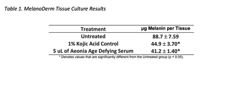


The purpose of this study was to assess the ability of the test material to reduce the melanin content of
Melanoderm tissues. In this study, a 5 ul topical application of the serum every 48 hours was observed
to significantly reduce tissue melanin content. Both the kojic acid and the serum treatment reduced
melanin in the quantitative assay when compared to the untreated group and this decrease in melanin
content is reflected in the lighter color of the melanocytes in the microscopic tissue images from these
two treatment groups when compared to the untreated group. In addition, treatment with the serum
was not observed to impact melanocyte morphology in this study. Photographic images of the tissues
demonstrate that the melanocytes from all three treatment groups display a normal, dendritic
morphology with the only difference between the treatment groups being the darkness of the
pigmentation.